Tissues in the shoot are derived from the shoot apical meristem (SAM). Just like in the root, the SAM produces three primary meristems, which produce the primary tissues:
Protoderm → Epidermis
Ground meristem → Cortex and pith (simply ground tissue in monocots)
Procambium → Primary xylem and primary phloem
These primary tissues will then either differentiate into specialized cells or, as is the case in many eudicots, become meristematic and produce secondary tissues.
Coleus Stem Tip
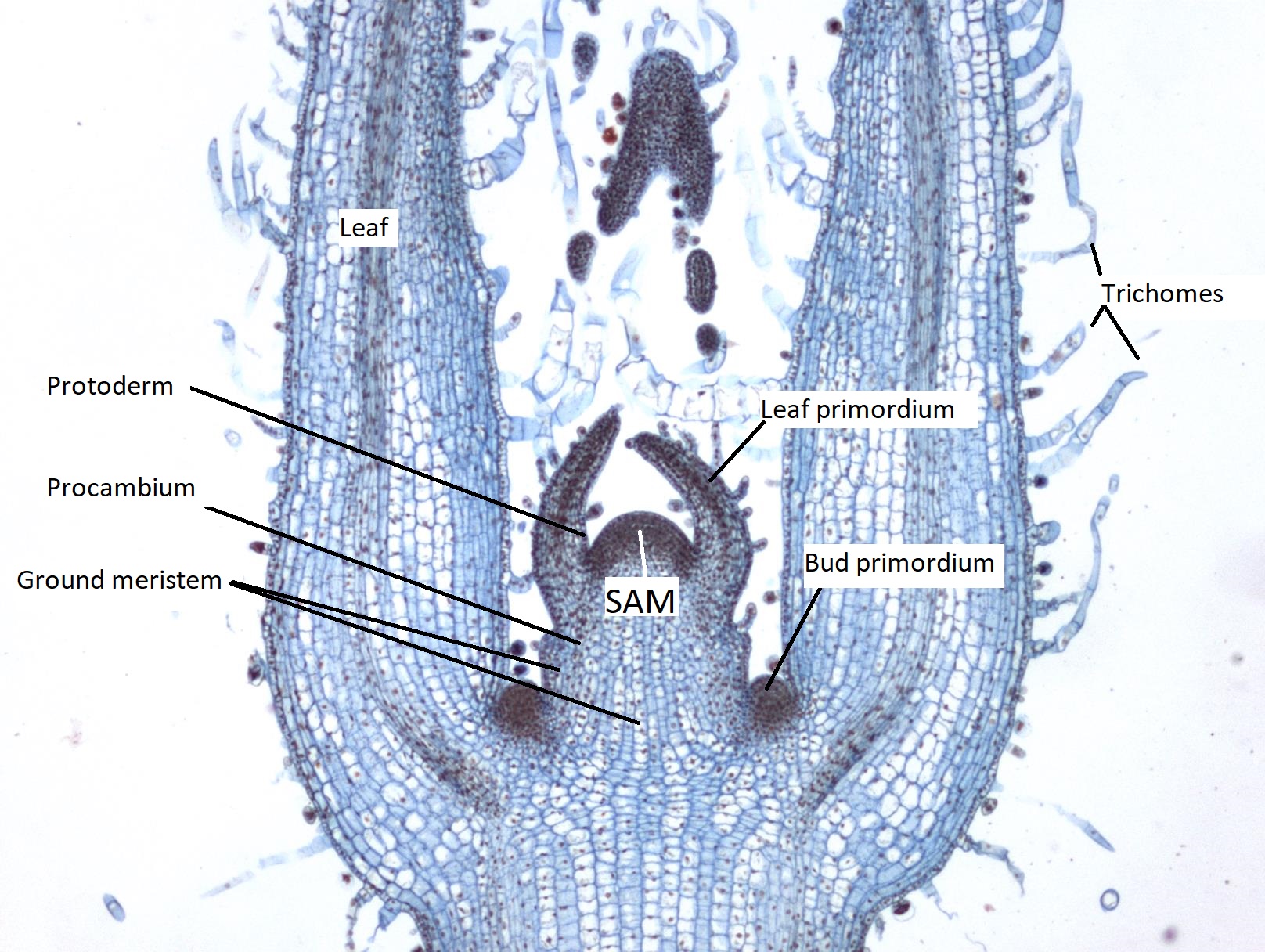
Though it looks a bit alien, this is a section through a growing tip of a plant. In the center, where the alien’s head might be, is a region of small, densely packed cells. This is the SAM of the apical bud. On either side of the SAM, like two upraised arms, are the leaf primordia. These are the early stages of developing leaves. Through the center of these leaf primordia is a darker region of small cells. This is the procambium, which will develop into the vascular tissue. Lining the outer edge of the SAM and the youngest portions of the leaf primordia is the protoderm. As the protoderm matures into the epidermis, it produces hair-like projections called trichomes. Between the protoderm and the procambium is the ground meristem.
On either side of the growing tip are two other darkened lumps of densely packed cells. These bud primordia will develop into axillary buds, producing either branches or flowers. Each bud primordium has its own SAM.
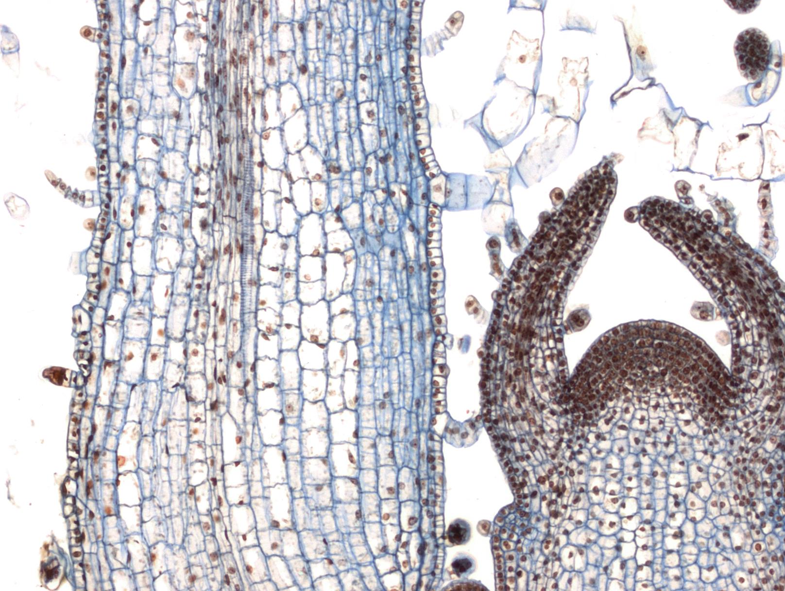
Figure \(\PageIndex{1}\): A partial view of a Coleus shoot tip long section. If we anthropomorphize it a bit and think of it as a humanoid with horns, it might be easier to conceptualize where the different parts are. The two "arms", held straight up into the air, are the leaves. Each leaf has many multicellular trichomes along its epidermis and a central strand of vascular tissue. Where the shoulders would be, there are two dark regions. These are bud primordia that will develop into axillary buds. They are dark in color because they are regions of intense cell division as they are rapidly growing. The "horns" are leaf primordia that will develop into leaves, much like the "arms". Between the two horns, at the top of the "head", is the shoot apical meristem (labeled SAM). All of the cells in the shoot are derived from this region. The protoderm runs along the exterior, near the SAM and leaf primordia. The procambium forms the vascular tissue and can be found in the center of the leaf primordia. The ground meristem forms the cortex and pith and can be found in the regions between the protoderm and procambium. Photo by Maria Morrow, CC BY-NC.Figure \(\PageIndex{2}\): A closer view of the Coleus shoot tip long section. Can you locate the SAM and primary meristems? Look for regions of densely packed cells, as these are all meristematic tissues, actively dividing to produce more cells. Photo by Maria Morrow, CC BY-NC.
Monocots
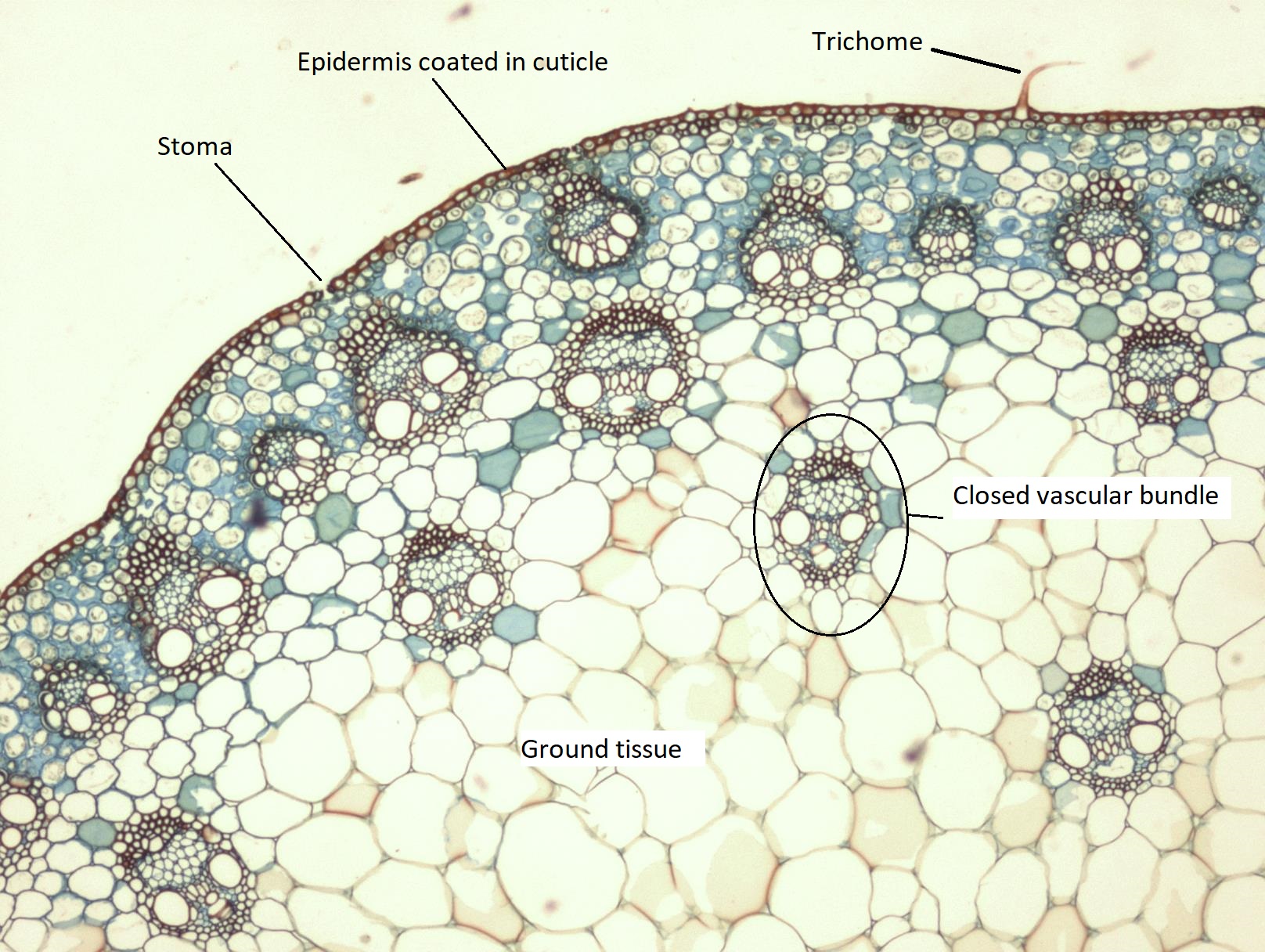
Figure \(\PageIndex{3}\): A cross section of a Zea mays stem. The organization of tissues differs from the Zea mays root. Most notably, the vascular tissue is arranged in bundles, rather than a central cylinder. These bundles are densely packed toward the outside of the stem, then occur less frequently toward the inside. Because there is no distinct delineation of the tissues produced by the ground meristem, it is now called ground tissue (as opposed to cortex and/or pith).Photo by Maria Morrow, CC BY-NC.
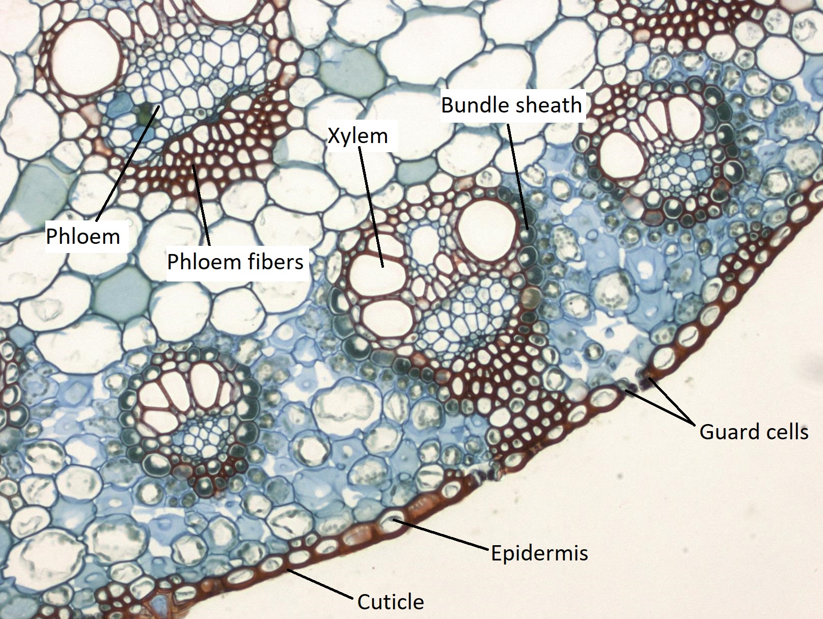
Figure \(\PageIndex{4}\): A closer view of a portion of the Zea mays stem. The epidermis is the outermost layer, coated in a thick cuticle that stains red. There are gaps in the epidermis where stomata are located, as well as a hair emerging (a trichome). The vascular bundles are situated within the ground tissue.Photo by Maria Morrow, CC BY-NC.Figure \(\PageIndex{5}\): A cross section of a Zea mays stem highlighting the vascular bundles. In the epidermis, the cuticle and guard cells are easier to distinguish in this image. Vascular bundles are arranged close to the epidermis with the xylem (stained red) toward the interior of the stem and the phloem (stained blue) toward the exterior. Each bundle is surrounded by a layer of cells called the bundle sheath. Photo by Maria Morrow, CC BY-NC.
Eudicots
Helianthus
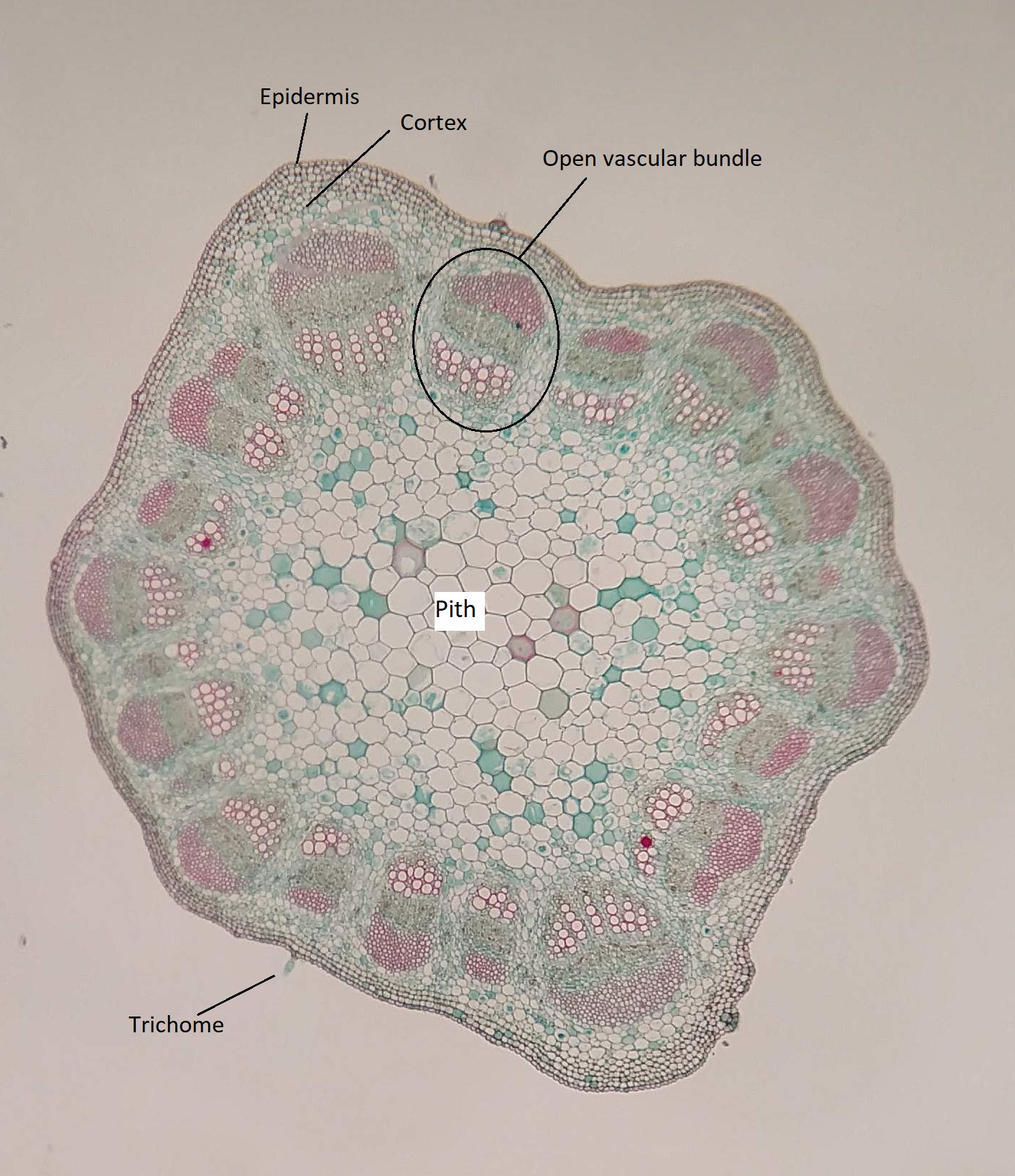
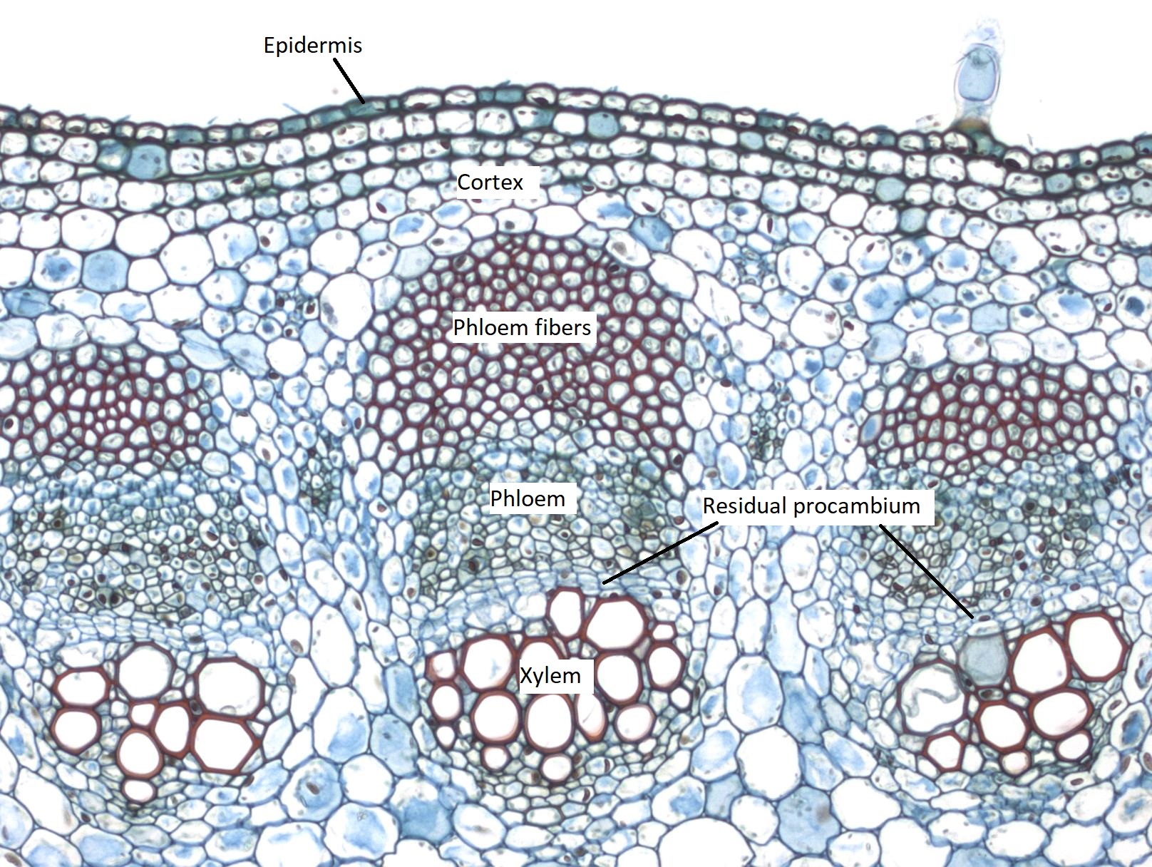
Figure \(\PageIndex{6}\): A cross section of a Helianthus stem. The outermost layer of cells is the epidermis. There is at least one trichome visible. Inside the epidermis is the cortex, which is composed largely of collenchyma in this plant. A ring of vascular bundles separates the cortex from the pith. Regions between the bundles are called pith rays. These vascular bundles are considered "open" because they have procambium tissue. Photo by Maria Morrow, CC BY-NC.Figure \(\PageIndex{7}\): A closer view of a young Helianthus stem. The outermost layer of cells (in this image, the topmost) is the epidermis. There is a blue, light-bulb shaped structure emerging from the epidermis in the upper left. This is a trichome. Beneath the epidermis, three layers of densely packed collenchyma cells and several layers of parenchyma make up the cortex. Three large vascular bundles are shown. There is a large cluster of phloem fibers at the top of each bundle, stained red due to the secondary walls. Below that is the phloem, which is separated from the xylem (large, open cells that are stained red) by the residiual procambium (densely packed, light blue cells).Photo by Maria Morrow, CC BY-NC.