Flowers are composed of many distinct components: sepals, petals, stamens, and carpels. These components are arranged in whorls and attach to an area called the receptacle, which is at the end of the stem that leads to the flower. This stem is called the peduncle. In the case of an inflorescence, where multiple florets are produced in place of a single flower, the stems leading to the florets are called pedicels.
Figure \(\PageIndex{1}\): This diagram shows a long section through a flower. Starting from the bottom, there is a stem called the peduncle. The peduncle terminates in a region called the receptacle, where all of the parts of the flower are attached. Sepals are found on the outside of the flower, two are visible here, with petals located just within the ring of sepals. There are five petals visible. Inside the petals, six stamens encircle a central pistil (composed of fused carpels). Diagram by Nikki Harris CC BY-NC with labels added.Figure \(\PageIndex{2}\): An image of an Allium inflorescense. Many small florets on stalks (pedicels) emerge from a central point at the tip of the peduncle. At this junction, papery leaves (bracts) can be seen. These features are labeled in a sketch shown to the right of the image. Photo by ramazan_murtazaliev, CC-BY-NC. Sketch by Maria Morrow, CC BY-NC.
Whorls
Flowers are composed of sets of highly modified leaves arranged in whorls. The outermost whorl of a flower is called the calyx and is composed of sepals. Inside the calyx is the corolla, which is composed of petals. The sepals are often smaller and less colorful than the petals, but this general rule can be misleading. For example, lilies often have identical sepals and petals. The only way you can distinguish between them is by location: Which whorl is on the outside?
Together, the calyx and corolla are called the perianth (peri- meaning around, anth- meaning flower).
The Perianth: Calyx and Corolla
Figure \(\PageIndex{3}\): A closed buttercup (Ranunculus sp.) flower shows the entire perianth. The sepals are smaller and covered in long trichomes. There are four sepals visible. Inside the sepal whorl (calyx), there are five overlapping, yellow petals. These petals form the corolla. Photo by Maria Morrow, CC BY-NC.Figure \(\PageIndex{4}\): Dissecting a flower is often necessary to see all parts of the flower. This floret has been cut in half and is being held down by a sharp probe. The sepals are short and pointed, the petals are longer and rounded. Because this is a floret, the stem leading up to it is called a pedicel instead of a peduncle. Photo by Melissa Ha, CC BY-NC.
Reproductive Parts: Androecium and Gynoecium
Figure \(\PageIndex{5}\): A dissected flower with all whorls labeled. The petals and sepals look the same, but can be distinguished by location. The stamens are similar in color to the perianth, located just inside the calyx. The gynoecium of this flower is located deep within a structure called a hypanthium, where all of the other floral whorls have fused together. The ovary is at the base of the hypanthium and is full of small, white ovules. Photo by Melissa Ha CC BY-NC with labels added.
Inside the perianth is the androecium (house of man), a whorl composed of stamens. Each stamen has a long filament holding up pollen sacs called anthers. Inside the androecium is the gynoecium (house of woman), which is composed of carpels. Each carpel has an ovary at the base where ovules are housed. The style emerges from the ovary and is topped by the stigma. Pollen grains land on the stigma and must grow a tube down the style to reach the ovule and complete fertilization.
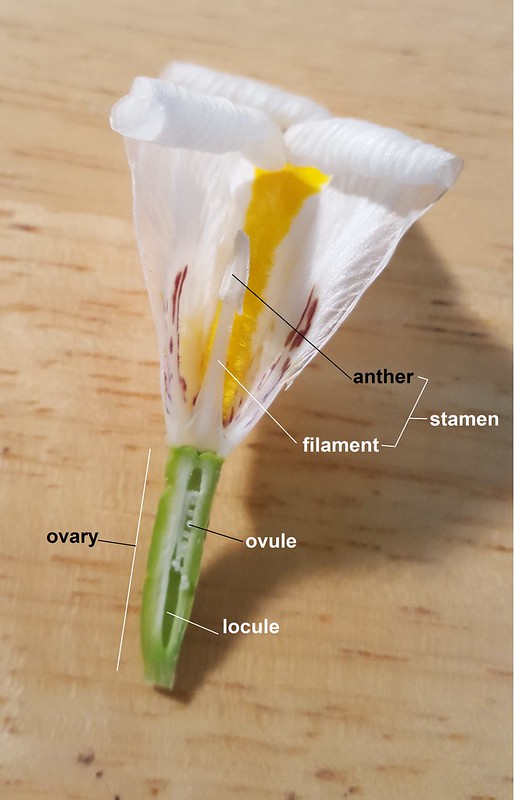
Figure \(\PageIndex{6}\): Here we see two sides of the same flower, which has been cut in half lengthwise. The first image shows the gynoecium, including two styles that fan out like wings toward the top where the stigma lobes are located. At the base of the styles, there is an elongated green ovary with many small white ovules inside. Each compartment of the ovary is called a locule. In the second image, the ovary is labeled, as well as the components of the androecium, including the anther and filament, which comprise the stamen. The anther is located at the end of the filament. Photos by Melissa Ha, CC BY-NC.
Androecium: The Stamen Whorl
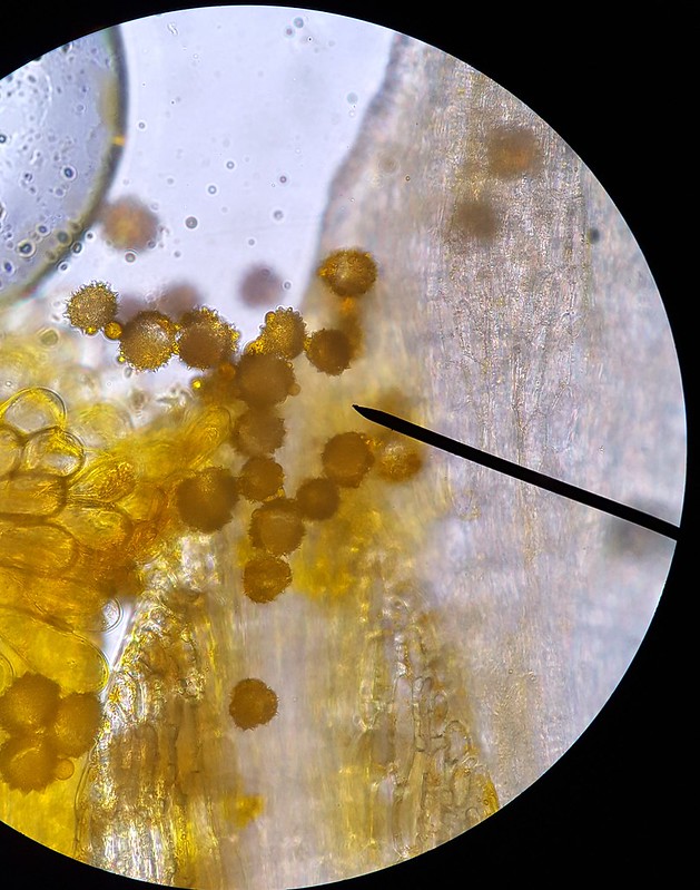
Figure \(\PageIndex{7}\): This buttercup has many stamens in the androecium. The anthers are flattened, long, and yellow. Yellow pollen grains produced within the anthers are scattered on the surface of an adjacent blade of grass. Photo by Maria Morrow, CC BY-NC.Figure \(\PageIndex{8}\): A single stamen that has been removed from a flower. The filament is long and flattened. At the end of the filament, there is an anther that has split partially open, releasing pollen grains. Photo by Melissa Ha, CC BY-NC .Figure \(\PageIndex{9}\): The image on the left shows two stigma lobes surrounded by anthers. The stigma lobes are covered in pollen. The image on the right shows the pollen at a higher magnification. The walls of the pollen are bumpy from ornamentation. Many species have characteristic pollen shapes and ornamentation that allow for identification of the plant species from the pollen grain, alone! Photos by Melissa Ha, CC BY-NC .
Gynoecium: The Carpel Whorl
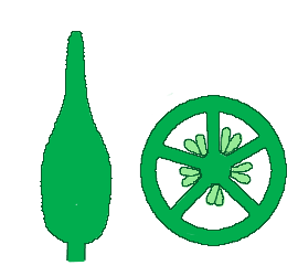
Figure \(\PageIndex{10}\): All of the whorls of this Trillium flower are visible in this picture. The sepals are on the exterior and green. The three petals are a light pink. There are six stamens, each with much longer anthers than filaments. The stamens encircle the enlarged ovary of this fertilized flower, which is grooved and beaked into an odd shape. A short style connects the ovary to three long, curling stigma lobes. Is this flower a monocot or eudicot? Photo by Maria Morrow, CC BY-NC.Figure \(\PageIndex{11}\): A syncarpous gynoecium in context. The gynoecium (whether composed of a single carpel or multiple "fused" carpels) is typically made up of an ovary, style, and stigma as in the center of the flower. Image by LadyofHats, Public domain, via Wikimedia Commons.Figure \(\PageIndex{12}\): In an apocarpous gynoecium, the carpels are not fused together. The illustrated gynoecium on the left has three free carpels. A cross section of the ovaries shows the ovules inside. The gynoecium shown in the center is syncarpous, with fusion of the ovaries. A cross section of the ovary shows three sets of ovules. The gynoecium on the right is fully syncarpous with fusion of all parts (stigma, style, and ovary). The ovary cross section is divided into five distinct locules. Images by Michael G. Simpson. Redrawn and color: User:RoRo, Public domain, via Wikimedia Commons.