Biology LibreTexts

6.1.2: Selaginella

  • Page ID
    35335
  • \( \newcommand{\vecs}[1]{\overset { \scriptstyle \rightharpoonup} {\mathbf{#1}} } \) \( \newcommand{\vecd}[1]{\overset{-\!-\!\rightharpoonup}{\vphantom{a}\smash {#1}}} \)\(\newcommand{\id}{\mathrm{id}}\) \( \newcommand{\Span}{\mathrm{span}}\) \( \newcommand{\kernel}{\mathrm{null}\,}\) \( \newcommand{\range}{\mathrm{range}\,}\) \( \newcommand{\RealPart}{\mathrm{Re}}\) \( \newcommand{\ImaginaryPart}{\mathrm{Im}}\) \( \newcommand{\Argument}{\mathrm{Arg}}\) \( \newcommand{\norm}[1]{\| #1 \|}\) \( \newcommand{\inner}[2]{\langle #1, #2 \rangle}\) \( \newcommand{\Span}{\mathrm{span}}\) \(\newcommand{\id}{\mathrm{id}}\) \( \newcommand{\Span}{\mathrm{span}}\) \( \newcommand{\kernel}{\mathrm{null}\,}\) \( \newcommand{\range}{\mathrm{range}\,}\) \( \newcommand{\RealPart}{\mathrm{Re}}\) \( \newcommand{\ImaginaryPart}{\mathrm{Im}}\) \( \newcommand{\Argument}{\mathrm{Arg}}\) \( \newcommand{\norm}[1]{\| #1 \|}\) \( \newcommand{\inner}[2]{\langle #1, #2 \rangle}\) \( \newcommand{\Span}{\mathrm{span}}\)\(\newcommand{\AA}{\unicode[.8,0]{x212B}}\)

    Members of the genus Selaginella are heterosporous, meaning they produce two different types of spores.
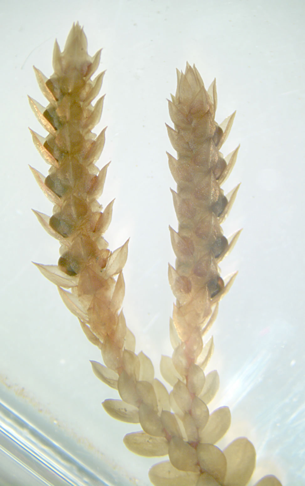
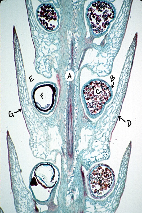
    Two Selaginella strobili with different looking sporangia A long section through a Selaginella strobilus showing microspores and megaspores
    Figure \(\PageIndex{1}\): In the first image, there are liquid-preserved strobili of Selaginella, showing mega- and microsporangia through translucent sporophylls. The second image is a cross section through a strobilus. Notice that the spores contained within the sporangia are quite different in size. Labels are as follows: A) cone axis, B) microsporangium, C) microspores, D) microsporophyll, E) megasporangium, F) megaspore, G) megasporophyll. Photo on the left is by Curtis Clark, licensed as noted, CC BY-SA 2.5, via Wikimedia Commons. Photo on the right is by Curtis Clark, CC BY-SA 3.0, via Wikimedia Commons with labels added by Maria Morrow.
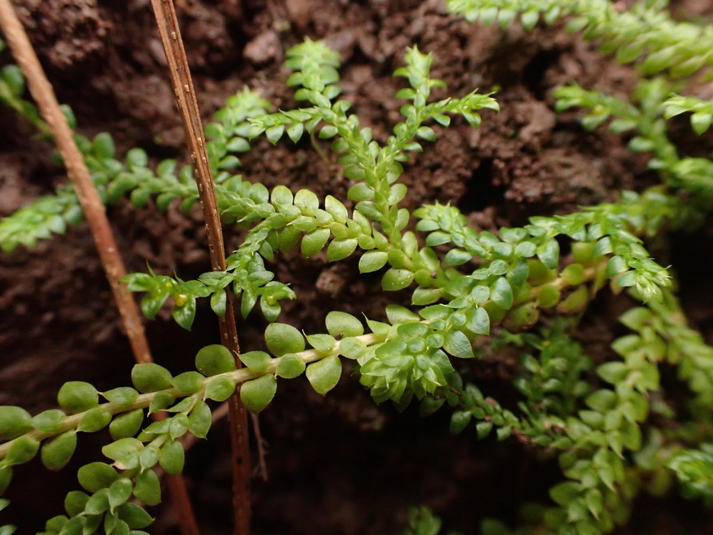
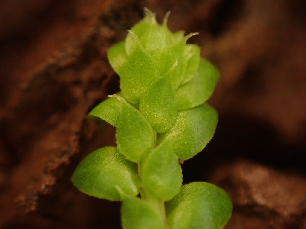
    Selaginella sporophyte spreading across the ground Close up of Selaginella microphylls
    Figure \(\PageIndex{2}\): Selaginella sporophytes have microphylls. These images show a Selaginella sporophyte spreading via dichotomous branching (first image) and the single, unbranched vein of vascular tissue present in each leaf (second image). Images by Sönke Bonde, CC0.

    This page titled 6.1.2: Selaginella is shared under a CC BY 4.0 license and was authored, remixed, and/or curated by Maria Morrow (ASCCC Open Educational Resources Initiative) .

    • Was this article helpful?