Biology LibreTexts

3.5.2: Types of Ascocarps

  • Page ID
    35312
  • \( \newcommand{\vecs}[1]{\overset { \scriptstyle \rightharpoonup} {\mathbf{#1}} } \) \( \newcommand{\vecd}[1]{\overset{-\!-\!\rightharpoonup}{\vphantom{a}\smash {#1}}} \)\(\newcommand{\id}{\mathrm{id}}\) \( \newcommand{\Span}{\mathrm{span}}\) \( \newcommand{\kernel}{\mathrm{null}\,}\) \( \newcommand{\range}{\mathrm{range}\,}\) \( \newcommand{\RealPart}{\mathrm{Re}}\) \( \newcommand{\ImaginaryPart}{\mathrm{Im}}\) \( \newcommand{\Argument}{\mathrm{Arg}}\) \( \newcommand{\norm}[1]{\| #1 \|}\) \( \newcommand{\inner}[2]{\langle #1, #2 \rangle}\) \( \newcommand{\Span}{\mathrm{span}}\) \(\newcommand{\id}{\mathrm{id}}\) \( \newcommand{\Span}{\mathrm{span}}\) \( \newcommand{\kernel}{\mathrm{null}\,}\) \( \newcommand{\range}{\mathrm{range}\,}\) \( \newcommand{\RealPart}{\mathrm{Re}}\) \( \newcommand{\ImaginaryPart}{\mathrm{Im}}\) \( \newcommand{\Argument}{\mathrm{Arg}}\) \( \newcommand{\norm}[1]{\| #1 \|}\) \( \newcommand{\inner}[2]{\langle #1, #2 \rangle}\) \( \newcommand{\Span}{\mathrm{span}}\)\(\newcommand{\AA}{\unicode[.8,0]{x212B}}\)
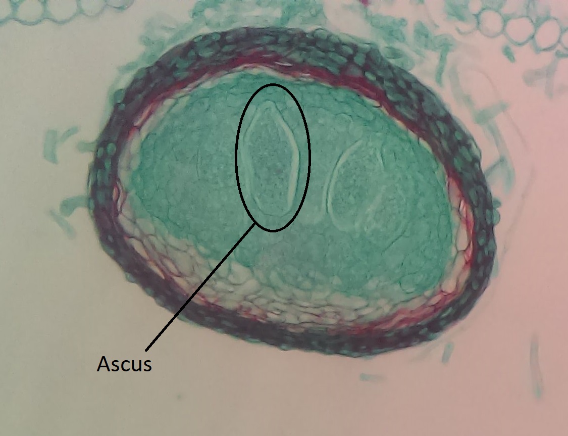
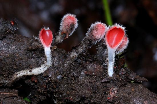
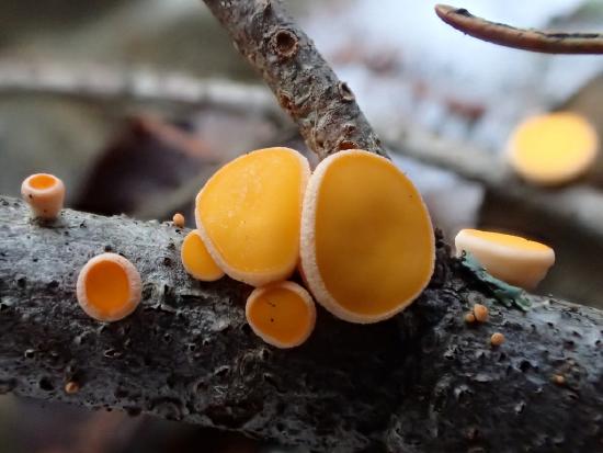
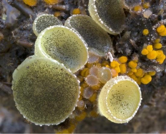
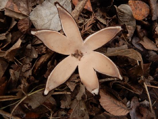
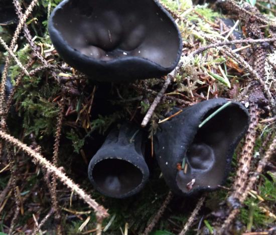
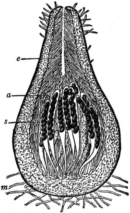
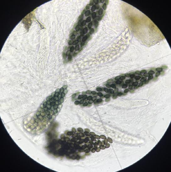
    Apothecium

    Apothecia are cup-shaped with the hymenium fully exposed (lining the interior of the cup), though this can be inverted and take on strange shapes (see Figure \(\PageIndex{3}\)).
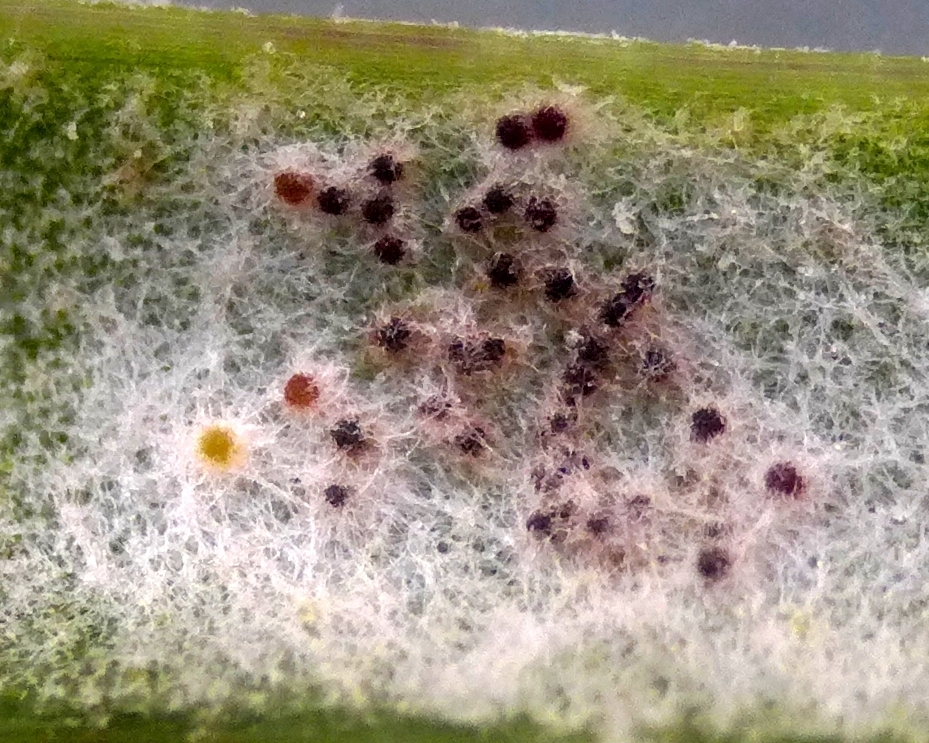
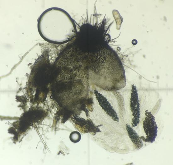
    A cluster of bright pink apothecia, almost fully closed, stalked, and covered in fuzzy white hairs
    Flat yellow disc-like apothecia growing on a branch
    Several yellow jelly discs with black asci visible
    A cup fungus that has split into a star-like shape
    Figure \(\PageIndex{1}\): A diversity of apothecia. Upper left: The stalked apothecium of Microstoma floccosum, photo by Alan Rockefeller. Upper right: The more classic disc-shaped apothecium of Pithya vulgaris, photo by Tyson Ehlers. Lower left: Gelantinous apothecia of Ascobolus furfuraceus, photo by Salvatore Bacciu and Paola Mereu. Lower right: The apothecium of Chlorioactis geaster splits into a star shape, photo by Bob O'Kennon. All photos licensed CC-BY-NC.
    Three apothecia with a more standard cup shape
    Figure \(\PageIndex{2}\): Three apothecia of the delightfully squishy Urnula padeniana. This type of fruiting body has a cup shape. The interior of the cup is where the spores are produced. Photo by Maria Morrow, CC-BY-NC.
    Two fruiting bodies with inverted apothecia held aloft on stalks Two fruiting bodies with a distinct stalk and an orange, tongue-like inverted apothecium
    Figure \(\PageIndex{3}\): Two apothecia of Helvella in the first image. This ascomycete, much like a morel, has an apothecium at the end of a stalk. The apothecium at the top of the stalk is not cup-shaped because the cup has been inverted. The spores are produced on the surface of the apothecium that you can see in this image. The second image shows another ascoymcete, Heyderia abietis, with a similar strategy. The inverted cup shape makes a smooth tongue-like structure on the top of the stalk. These are only about 1 cm tall. Photos by Maria Morrow, CC-BY-NC.

    Perithecium

    A flask-shaped fruiting structures, often microscopic and/or embedded within either the substrate it is fruiting in or a fungal structure called a stroma.
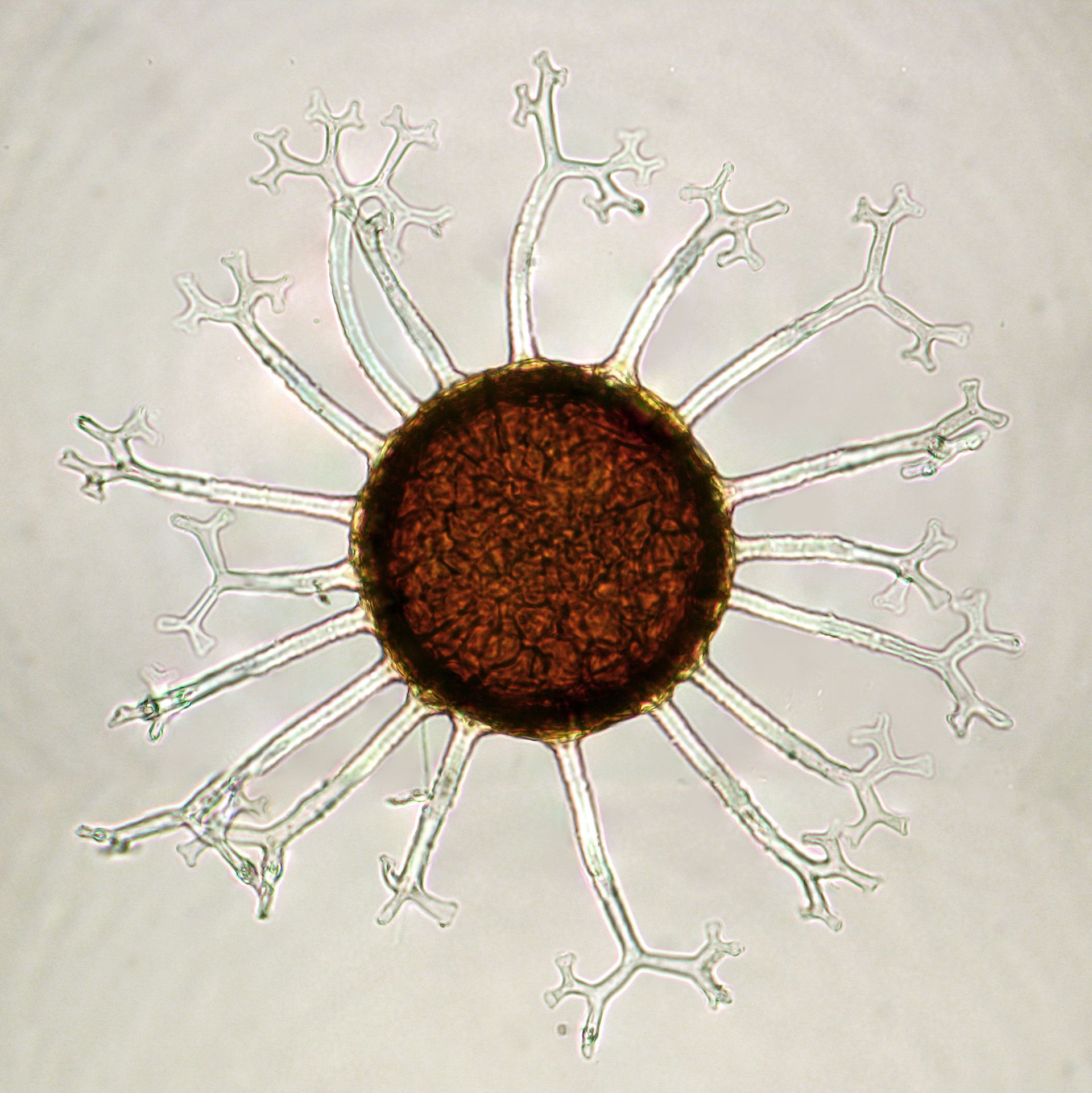
    Long section through a perithecium
    Figure \(\PageIndex{4}\): A flask-shaped perithecium, with asci emerging within the base of the flask. Labels in the diagram are as follows: m=the mycelium, s=asci with black ascospores inside, a=paraphyses (sterile cells surrounding the asci), e=the ostiole (opening). From Strasburger’s Lehrbuch der Botanik. After v. Tavel., Public domain, via Wikimedia Commons.
    A perithecium that has been squished to reveal the asci Asci from the perithecium in the previous picture. Each large ascus contains 20+ ascospores.
    Figure \(\PageIndex{5}\): A Podospora perithecium viewed under the microscope. Short hairs surround the ostiole. It has been squished a bit and the asci have erupted out the base. Unlike many ascomycetes, many spores are produced inside the asci of this species. Photos by Sigrid Jakob, CC-BY-NC.
    A single, hairy perithecium sits atop its substrate At least two hairy perithecia are visible here
    Figure \(\PageIndex{6}\): Some perithecial fungi, like the Chaetomium sp. pictured here, form their perithecia on the surface of whatever substrate they are growing on. Look for the small, hairy, black, pear-shaped fruiting bodies in the images above. Photos by Photos by Sigrid Jakob, CC-BY-NC.
    fruiting structure of Xylaria. There is a skinny black stalk, then the top half is covered by large, round lumps. A long section through the Xylaria fruiting structure. The internal tissue is white and surrounds black cavities (the perithecia).
    Figure \(\PageIndex{7}\): Xylaria species produce perithecia embedded in sterile fungal tissue called a stroma. In Xylaria tucumanensis, the perithecia are large and are not surrounded by much stroma. In the first photo (left), each lumpy projection has a distinct dot in its center; this is the ostiole (opening) of the perithecium. The second photo (right) shows a long section through a fruiting structure. The white tissue is the stroma. The black, concave structures are the perithecia. An arrow points to an ostiole of one of the perithecia. Photos by Danny Newman, CC-BY-NC-SA.

    Cleistothecium

    A cleistothecium is a fully-enclosed fruiting structure. These typically have bag-like asci. Some split open to release their spores and are called chasmothecia.

    A section through a cleistothecium with an ascus circled
    Figure \(\PageIndex{8}\): A section through a cleistothecium with an ascus circled. A cleistothecium is a fully enclosed fruiting body, generally microscopic, that encloses many bag-like asci. Photo by Melissa Ha, CC-BY-NC with labels added.
    A closed, spherical orange fruiting body with transparent, branching structures emerging from it
    Figure \(\PageIndex{9}\): Some cleistothecia, such as those of the powdery mildews (in the order Erysiphales), are beautifully ornamented. Gubin Olexander, CC BY 4.0, via Wikimedia Commons.
    A magnified view of some mycelium on a leaf, showing small spherical stuctures embedded in it.
    Figure \(\PageIndex{10}\): Powdery mildews (Erysiphales) are plant parasites that form cleistothecia. They get their name from the powdery white look they give to the surface of plants they are parasitizing (first photo). Sometimes within this powdery white fluff (mycelium), cleistothecia can be seen with some magnification (second photo). Photos by ðejay (Orkney), CC-BY-NC.

    This page titled 3.5.2: Types of Ascocarps is shared under a CC BY-NC 4.0 license and was authored, remixed, and/or curated by Maria Morrow (ASCCC Open Educational Resources Initiative) .

    • Was this article helpful?