Biology LibreTexts

3.5.1: Characteristics

  • Page ID
    35311
  • \( \newcommand{\vecs}[1]{\overset { \scriptstyle \rightharpoonup} {\mathbf{#1}} } \) \( \newcommand{\vecd}[1]{\overset{-\!-\!\rightharpoonup}{\vphantom{a}\smash {#1}}} \)\(\newcommand{\id}{\mathrm{id}}\) \( \newcommand{\Span}{\mathrm{span}}\) \( \newcommand{\kernel}{\mathrm{null}\,}\) \( \newcommand{\range}{\mathrm{range}\,}\) \( \newcommand{\RealPart}{\mathrm{Re}}\) \( \newcommand{\ImaginaryPart}{\mathrm{Im}}\) \( \newcommand{\Argument}{\mathrm{Arg}}\) \( \newcommand{\norm}[1]{\| #1 \|}\) \( \newcommand{\inner}[2]{\langle #1, #2 \rangle}\) \( \newcommand{\Span}{\mathrm{span}}\) \(\newcommand{\id}{\mathrm{id}}\) \( \newcommand{\Span}{\mathrm{span}}\) \( \newcommand{\kernel}{\mathrm{null}\,}\) \( \newcommand{\range}{\mathrm{range}\,}\) \( \newcommand{\RealPart}{\mathrm{Re}}\) \( \newcommand{\ImaginaryPart}{\mathrm{Im}}\) \( \newcommand{\Argument}{\mathrm{Arg}}\) \( \newcommand{\norm}[1]{\| #1 \|}\) \( \newcommand{\inner}[2]{\langle #1, #2 \rangle}\) \( \newcommand{\Span}{\mathrm{span}}\)\(\newcommand{\AA}{\unicode[.8,0]{x212B}}\)
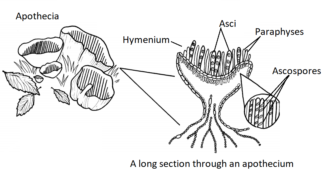
    An apothecium and a long section through an apothecium
    Figure \(\PageIndex{1}\): Ascomycetes are called the cup fungi after the cup-shaped fruiting body formed by many of the larger organisms in this group, the apothecium. A few apothecia are shown on the left side of the diagram. The right side shows a long section through an apothecium. Septate hyphae comprise most of the structure. On the interior surface of the cup is the hymenium, which is composed of asci and paraphyses. Ascospores are produced by meiosis within each ascus. Diagram by Nikki Harris, CC-BY-NC, with labels added by Maria Morrow.
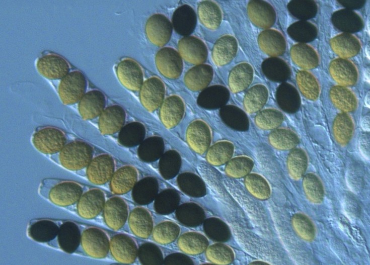
    Elongate, transparent sacs filled with spores, half dark and half light.
    Figure \(\PageIndex{2}\): In this photo, light and dark colored ascospores are produced inside the asci of the fungus Sordaria macrospora. This demonstrates the linear division of the spores, as pairs of genetically identical light or dark spores are adjacent, produced after mitosis occurs within the ascus. Photo by Aurora Storlazzi, CC BY 4.0, via Wikimedia Commons.
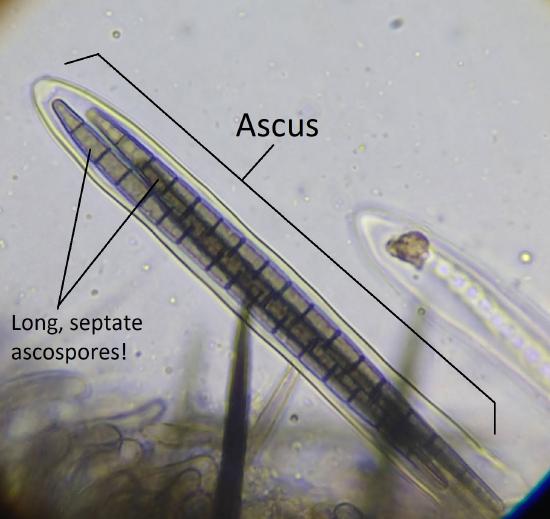
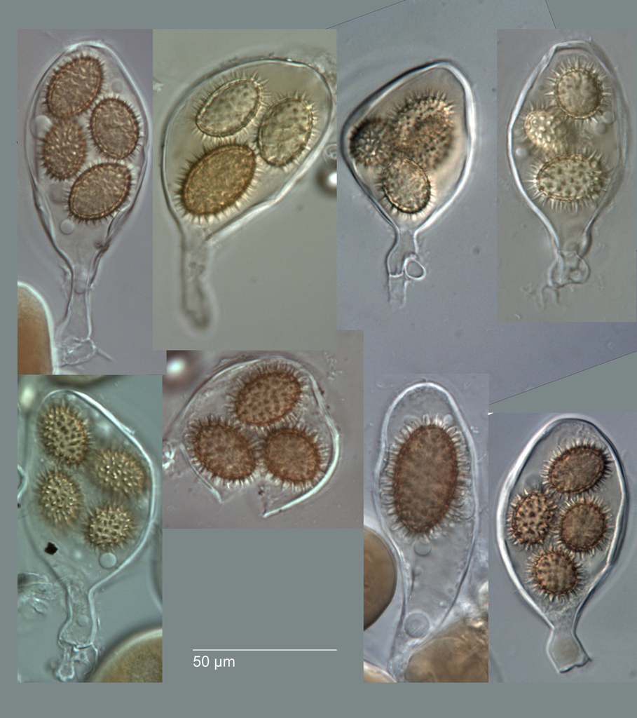
    An ascus with several long, septate ascospores inside it Multiple, bag-like asci. Each contains 3-4 huge, spiky spores.
    Figure \(\PageIndex{3}\): Ascospores and asci can have an incredible diversity of sizes, shapes, and structure. In the first photo (left), a single ascus surrounds several ascospores (approximately three can be distinguished, here). Each ascospore is as long as the ascus and looks striped. These stripes are septations and each spore has many septa. In the next photo (right), 3-4 enormous spiny spores of Tuber rufum are produced in wider, bag-like asci. First photo by Maria Morrow, CC-BY-NC. Second photo by Jerry Cooper, CC-BY.
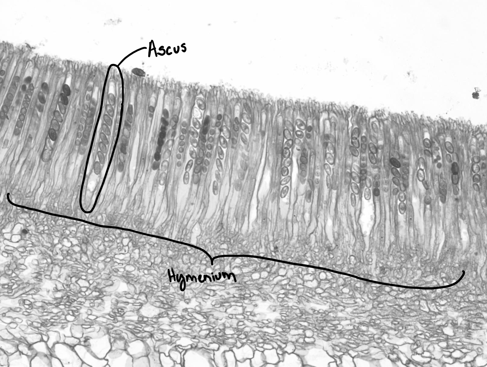
    A prepared slide of the hymenium of a cup fungus, showing a row of asci with ascospores (this row is labeled as the hymenium)
    Figure \(\PageIndex{4}\): Asci, some filled with ascospores, lining the hymenium of a Peziza fruiting body. Photo by Maria Morrow, CC-BY-NC.
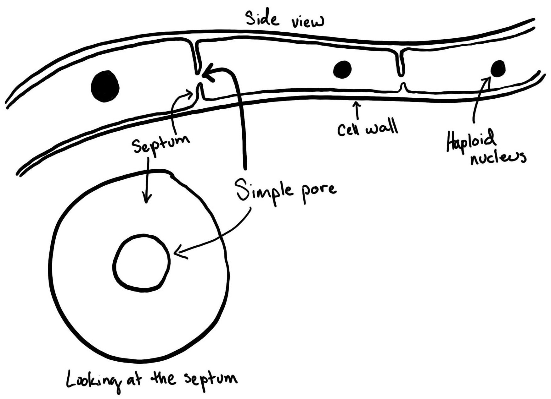
    A hyphal filament with simple septations. Each septum has a hole in the center, looking like a donut from a straight-on view.
    Figure \(\PageIndex{5}\): Ascomycetes have septate hyphae with a simple pore (hole) in the center. This separates hyphae into distinct cells yet still allows for the passage of materials between these compartments. This drawing shows a side view of a hypha with two septations drawn. Each septation has a gap in the center. Below, a septum is shown as if you were looking straight at it, showing that it is a ring of tissue with a hole in the center. Each compartment of the hypha has a single haploid nucleus. Drawing by Maria Morrow, CC-BY-NC.

    This page titled 3.5.1: Characteristics is shared under a CC BY-NC 4.0 license and was authored, remixed, and/or curated by Maria Morrow (ASCCC Open Educational Resources Initiative) .

    • Was this article helpful?