
7.4: Chromosomes and Karyotypes
- Page ID
- 24772

Chromosomal Features
![By NIH, User:Phrood, En rouge [Public domain or Public domain], via Wikimedia Commons](https://bio.libretexts.org/@api/deki/files/36623/chromosomes-953x1024.png?revision=1&size=bestfit&width=612&height=658)
Chromosomes are made of double-stranded DNA molecules wound about histones and condensed into the familiar X-shape. Under regular functioning, these chromosomes are decondensed in the nucleus and not recognizable.
Chromosomes in Interphase are not visible individually. In preparation for nuclear division (mitosis or meiosis), they begin to organize tighter and condense in preparation for movement to subsequent daughter nuclei. The animation below illustrates the process of histone packaging and the molecular visualization of DNA replication. Histones are proteins that aid in the packaging of the chromosomes into organized coils that give rise to the recognizable chromosomes during metaphase.
Large-scale genomic rearrangements result in genetic abnormalities. Biologists utilize a technique called a chromosome spread followed by a karyotype or karyogram. To make a chromosome spread, one blocks the progression of mitosis at metaphase where chromosomes are condensed into the structures we are familiar with. A karyotype analysis is an arrangement of the chromosome spread into the homologous pairs of chromosomes.
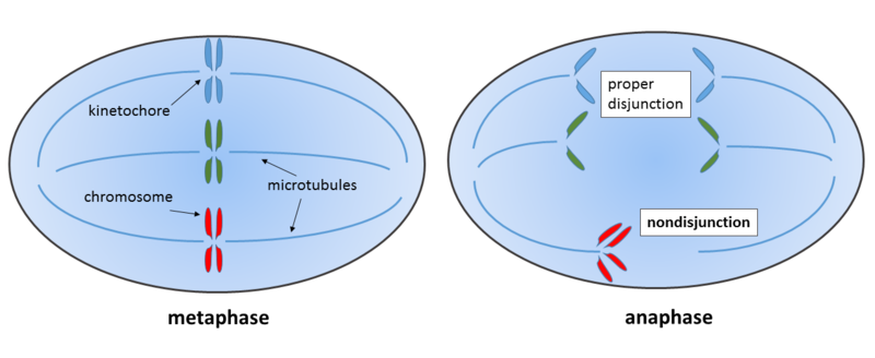
Events associated with the improper separation of chromosomes during metaphase results in an alteration of chromosome number in the subsequent generation of cells. Using the Pop-beads, we can understand better how the timing of these events will lead to differences in the karyotype.
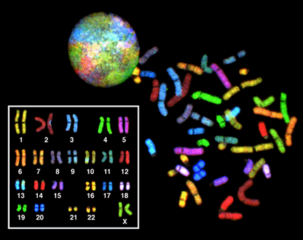
A “spectral” karyotype of a female nucleus. Each homologous pair is “painted” to differentiate them.
Abnormal Karyotypes
Down’s Syndrome is a common genetic abnormality referred to as Trisomy 21. Instead of having the complement of 46 chromosomes of 22 homologous pairs plus 2 sex chromosomes, there are 47 chromosomes consisting of an additional Chromosome 21.
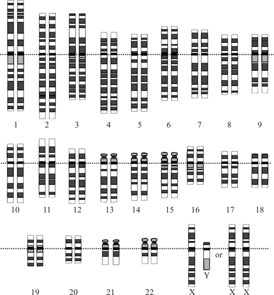
Standard Human Karyotype with 46 chromosomes. Both XX and XY are also shown here.
The appearance of extra or missing chromosomes arises during meiosis in an event called nondisjunction. After fertilization, a zygote with an improper chromosome complement occurs.

From left to right, the ploidy of the resultant zygotes: 2N, 2N, 2N+1, 2N-1; 2N+1, 2N+1, 2N-1, 2N-1
Nondisjunction can occur during Meiosis I or Meiosis II to yield aneuploid states of 2N+1 or 2N-1.

Down’s syndrome karyotype showing the traditional trisomy 21.
Translocation
Translocation is the movement of a piece or a whole chromosome onto another chromosome. The acrocentric nature of Chr 21 and it’s small size makes it prone to an event called Robertsonian Translocation whereby two acrocentric chromosomes fuse.

Down’s Syndrome from a 14:21 translocation.
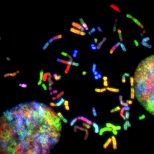
A spectral karyogram of a brain cancer (glioblastoma) illustrating multiple translocation problems and chromosome instability. Credit: Thomas Reid, NCI (CC-BY-NC)
Abnormalities in mitosis also occur and can result in diseases from translocations.

Polyploidy
Some organisms and cells have entire sets of chromosomes additional to the standard 2N diploid. Cells that have extra sets in the formula of 3N are called triploid. If they are 4N, they are called tetraploid. This is different than the case of Down’s syndrome, which has a chromosome complement of 2N+1. Any time there are abnormal numbers of chromosomes, cells are referred to as aneuploid. A special case of aneuploid occurs from having entire sets more of chromosomes—polyploid. Plants are especially robust in the regard of polyploidy and often have different species arise in such a way. Some plants become sterile in the case of polyploidy and will not produce seeds properly.

Wild bananas or plantains (Musa acuminata or Musa balbisiana) are deemed inedible because of their large seeds.
Have you ever seen a banana with these large seeds? The answer is most likely “No!” since these are not regarded as being edible. However, due to selective breeding practices, most edible plantains and bananas are hybrids of the two species Musa acuminata or Musa balbisiana that are 3N or 4N. In this case, the fruits are sterile and the seeds don’t develop. Other seedless fruits are also developed this way and require propagation through clonal means.
Advanced View of Chromosome Segregation