
Mitosis and Meiosis
- Page ID
- 2856
\( \newcommand{\vecs}[1]{\overset { \scriptstyle \rightharpoonup} {\mathbf{#1}} } \) \( \newcommand{\vecd}[1]{\overset{-\!-\!\rightharpoonup}{\vphantom{a}\smash {#1}}} \)\(\newcommand{\id}{\mathrm{id}}\) \( \newcommand{\Span}{\mathrm{span}}\) \( \newcommand{\kernel}{\mathrm{null}\,}\) \( \newcommand{\range}{\mathrm{range}\,}\) \( \newcommand{\RealPart}{\mathrm{Re}}\) \( \newcommand{\ImaginaryPart}{\mathrm{Im}}\) \( \newcommand{\Argument}{\mathrm{Arg}}\) \( \newcommand{\norm}[1]{\| #1 \|}\) \( \newcommand{\inner}[2]{\langle #1, #2 \rangle}\) \( \newcommand{\Span}{\mathrm{span}}\) \(\newcommand{\id}{\mathrm{id}}\) \( \newcommand{\Span}{\mathrm{span}}\) \( \newcommand{\kernel}{\mathrm{null}\,}\) \( \newcommand{\range}{\mathrm{range}\,}\) \( \newcommand{\RealPart}{\mathrm{Re}}\) \( \newcommand{\ImaginaryPart}{\mathrm{Im}}\) \( \newcommand{\Argument}{\mathrm{Arg}}\) \( \newcommand{\norm}[1]{\| #1 \|}\) \( \newcommand{\inner}[2]{\langle #1, #2 \rangle}\) \( \newcommand{\Span}{\mathrm{span}}\)\(\newcommand{\AA}{\unicode[.8,0]{x212B}}\)
Mitosis in Animals
Human Chromosomes
- View a slide of human chromosomes and draw some of the chromosomes in your notebook.
- Do the chromosomes have one chromatid or two?
Below: Human Chromosomes Click on the photograph to view an enlargement.
Whitefish Blastula
- The cells of a developing embryo are dividing rapidly and can be used for viewing the different stages of mitosis. Obtain a whitefish blastula (early embryo) slide and find a cell in each of these phases: interphase, prophase, metaphase, anaphase, and telophase.
- Draw a cell in anaphase.
Mitosis in Plants
- Cells at the tips of plant roots and stems grow rapidly and can be used for viewing the stages of mitosis. Used a slide of onion (Allium) root tip to identify interphase, prophase, metaphase, anaphase, and telophase.
- Find a cell in telophase and draw the cell in your notebook. Draw the cell plate if it is visible.
Meiosis in Animals (Gametogenesis)
Testis
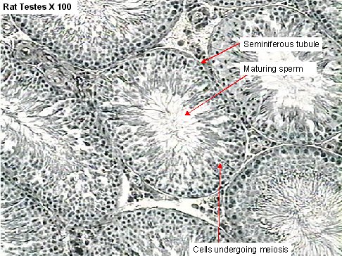
- Obtain a slide of a cross section of cat testes and observe the seminiferous tubules. Identify spermatogonia. Identify sperm.
Below: Rat testes X 100. Click on the image to view an enlargement.
Ovary
The primary oocyte is contained within a structure called a follicle. As the follicle enlarges, it produces hormones. During ovulation, the follicle ruptures and releases the secondary oocyte.
- View a slide of a section of a rabbit ovary under scanning magnification and observe follicles in various stages of development. Can you see an oocyte in any of the follicles?
Below: Rabbit ovary X 40. Click on the image to view an enlargement.
